WHY THIS MATTERS IN BRIEF
3D printing human organs on demand is one thing, but now 3D printed tumours can help researchers develop better cancer cures faster.
 Interested in the Exponential Future? Connect, download a free E-Book, watch a keynote, or browse my blog.
So far we’ve 3D printed everything from buildings, cars, and electronics, to human tissue and robots, and now researchers in the US have 3D printed cancers which might sound odd, but bear with me. In the US the cancer death rate has dropped 27 percent in the last quarter-century, resulting in just over 600,000 deaths from the disease nationally in 2019, and this steady decline is in large part thanks to new, effective cancer treatments that have transformed the disease from a death sentence to a survivable, chronic condition.
But despite these promising statistics, some cancers still resist traditional treatment. In particular, a type of aggressive, fast-growing brain tumor called a glioblastoma is resistant to many forms of cancer treatment and still has an expected survival rate of only 11 – 15 months.
Part of the challenge when it comes to treating tumors like glioblastomas, which can develop resistance to typical cancer drugs, is to determine how different treatments will affect the tumor growth before applying those treatments to cancer patients. The gold standard for this kind of testing is still often animal models, such as mice and rats, but these models can be tricky to work with and vary widely, making them non-ideal for repeated, controlled testing.
So, instead of relying on imperfect, biological samples to solve this problem, a multidisciplinary team of scientists decided to 3D print their own brain tumors instead.
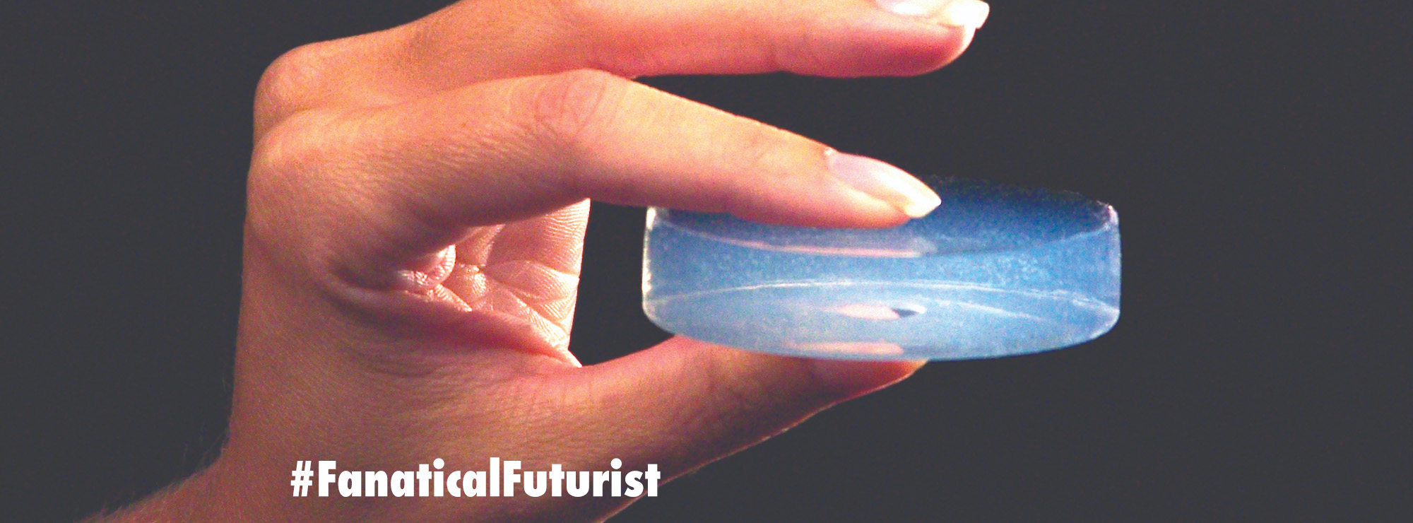
Using glioblastoma cancer cells derived from patients themselves, the team created a bio-ink that could be printed using a 3D bio-printer to create little, lumpy spheroids, or 3D tumors. Xavier Intes, co-author and co-director of project from Rensselaer Polytechnic Institute, told reporters these life-like models can help scientists better understand how to tackle this problem in a controlled environment.
“There’s a lot of drive to better understand the biology of tumors, and glioblastomas are among the most lethal ones,” said Intes. “They can vary very much patient to patient. One of the issues has always been understanding how we can try to tackle this disease. The unique aspect of this bioprinting is that it’s really going to print to biology.”
By printing the biology the researchers are then able to study their biological reaction to drugs in a controlled environment. Importantly, these tumors were even complete with bio-printed blood vessels for the cancer drugs to be delivered through. Intes went on to add that these vascular pathways are not only essential for creating a pathway for the drug to reach the cells but to also more accurately recreate a real tumor environment.
While vascular systems have been included in previous model studies, such as Tumors-on-a-Chip, this study is unique in applying them to 3D printed tumors.
Researchers were able to conduct their drug trials on the 3D tumor over 70 days, much longer than other non-living models, and were able to closely watch how the tumor responded to treatment using a new non-damaging imaging method designed for the trial.
The team designed a fast, deep-tissue imaging technique that could see through the thick Plexiglas container holding the tumors and down to their cellular activity. The speed of this approach allowed the researchers to take measurements with minimal photo damage to the tissue, the authors write. This allowed them to more accurately see if the drug had reached the cancer cells as well as if the cancer cells were dying as a result.
“We developed a new technology that allows us to go deeper than fluorescence microscopy,” Intes said in a statement. “It allows us to see, first, if the cells are growing, and then, if they respond to the drug.”
Intes said that this approach can not only be used to better understand how different therapies affect glioblastomas but can be applied to other cancers, such as breast and prostate, as well as other diseases at large.
Going forward, Intes says the team will work to improve the speed required to create and test these tumors so that life-saving information can reach the patients as soon as possible.