WHY THIS MATTERS IN BRIEF
Particle accelerators that fit on a chip will one day revolutionize medicine but in the meantime they’re transforming X-Rays.
 Love the Exponential Future? Join our XPotential Community, future proof yourself with courses from XPotential University, read about exponential tech and trends, connect, watch a keynote, or browse my blog.
In our desire to see and explore the human body in greater depth and detail than ever before recently we’ve seen the development of everything from small portable MRI and at home X-Ray machines, which can be wheeled to a patient’s bedside, all the way through to new revolutionary medical imaging technologies that let you video human cells in vivo in real time, and see the human body live in extreme detail. And then there are particle accelerators that fit on a chip … but that’s another story.
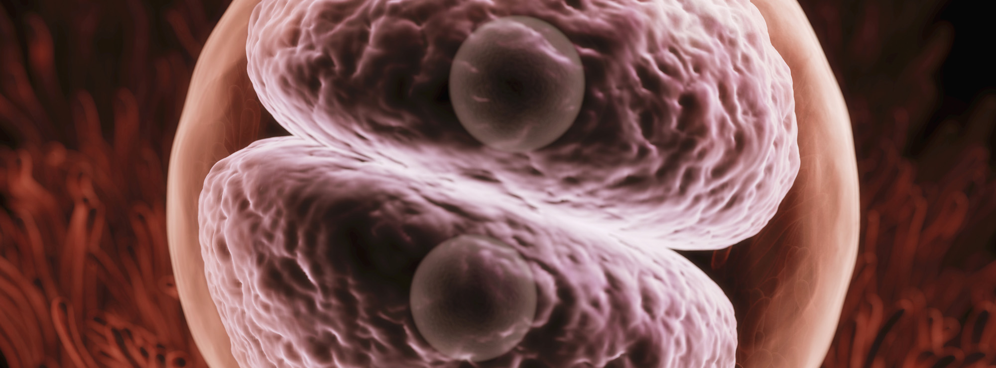
Now, adding to those advances, a groundbreaking new imaging technique, utilizing X-rays generated by a cutting-edge particle accelerator, is offering 3D images of whole organs in unprecedented detail. Demonstrating the technology researchers imaged the lung of a deceased COVID-19 patient, revealing novel insights into how the disease disrupts blood oxygenation.
learn about the Future of Healthcare, by Keynote Speaker Matthew Griffin
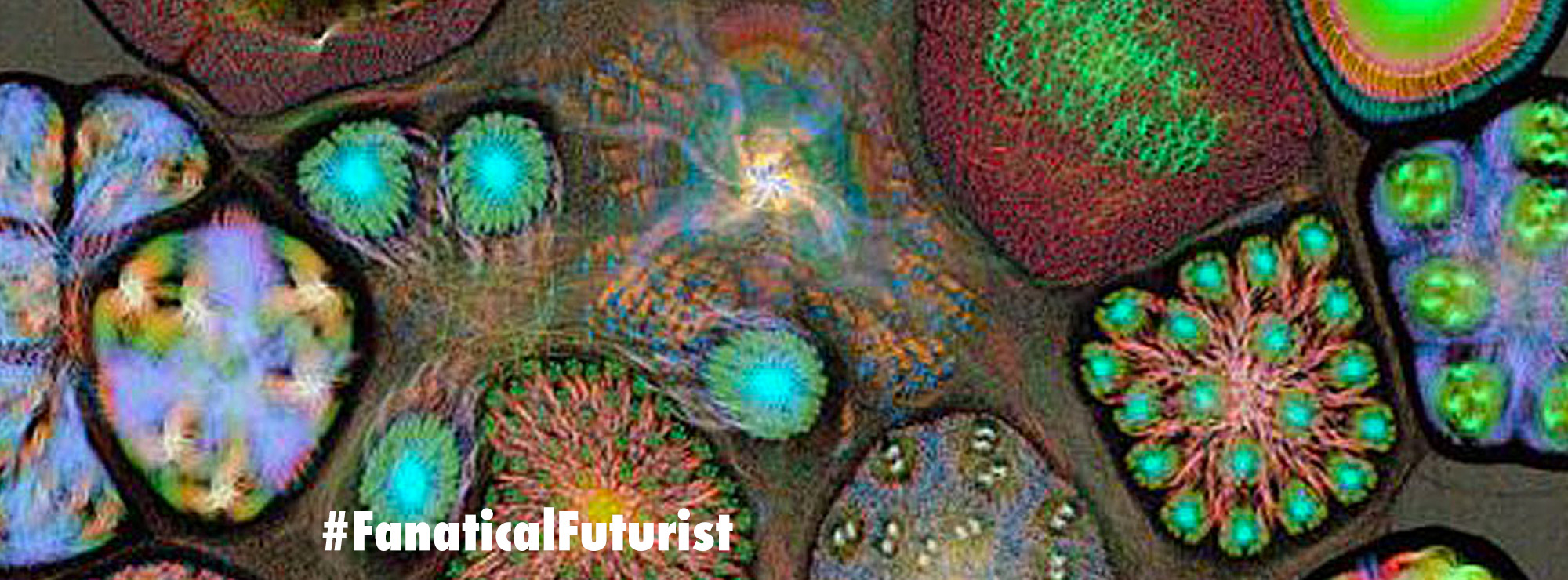
The new technology is called Hierarchical Phase-Contrast Tomography (HiP-CT) and is an X-ray technique that allows whole organs to be imaged down to a resolution of 1 micron – 100 times the resolution of a conventional CT scan.
The imaging advance comes from a technological upgrade at the European Synchrotron Research Facility (ESRF). This cutting-edge particle accelerator was improved recently with what is dubbed the Extremely Brilliant Source upgrade (ESRF-EBS).
This EBS upgrade created the world’s first fourth-generation synchrotron, making it the brightest X-ray source in the world. This increased X-ray performance by a factor of 100 in terms of “brilliance and coherence.” And the X-rays generated by this device are 100 billion times brighter than what is found in a conventional hospital X-ray.
X-Rays like you’ve never seen them before
“The idea to develop this new HiP-CT technique came after the beginning of the global pandemic, by combining several techniques that were used at the ESRF to image large fossils, and using the increased sensitivity of the new Extremely Brilliant Source at the ESRF, ESRF-EBS,” explains lead scientist at ESRF Paul Tafforeau, adding “This allows us to see in 3D the incredibly small vessels within a complete human organ, enabling us to distinguish in 3D a blood vessel from the surrounding tissue, and even to observe some specific cells.”
Using the new technology, a team led by researchers from University College London is launching a project called the Human Organ Atlas which complements the Human Cell Atlas that I talked about a while ago. Peter Lee, who is leading the project, says the goal of the Human Organ Atlas is to fill a gap in our understanding of human anatomy.
… see even more!
“Clinical CT and MRI scans can resolve down to just below a millimeter, whilst histology (studying cells/biopsy slices under a microscope), electron microscopy (which uses an electron beam to generate images) and other similar techniques resolve structures with sub-micron accuracy, but only on small biopsies of tissue from an organ,” says Lee. “HiP-CT bridges these scales in 3D, imaging whole organs to provide new insights into our biological makeup.”
The Human Organ Atlas will be a free online resource and it launches with displays of several key human organs, including the brain, kidneys, heart and spleen. The project also offers imaging comparing a healthy lung against a lung from a deceased COVID-19 patient.
A fundamental pathological sign of a COVID-19 deterioration is a sharp drop in blood oxygenation levels and HiP-CT imaging has revealed insights into how this occurs through a process known as “shunting.” It had been previously hypothesized that COVID-19 reduces blood oxygen rates by increasing levels of shunting in the lungs but this is the first direct evidence of that process.
“By combining our molecular methods with the HiP-CT multiscale imaging in lungs affected by COVID-19 pneumonia, we gained a new understanding [of] how shunting between blood vessels in a lung’s two vascular systems occurs in COVID-19 injured lungs, and the impact it has on oxygen levels in our circulatory system,” says Danny Jonigk, a researcher from Hannover Medical School working on the project.
HiP-CT is designed to offer doctors a library of images documenting how different diseases affect a variety of organs. This never-before-seen structural data illustrates how disease can influence tissue architecture down to resolutions as small as one micron.
Claire Walsh, a mechanical engineer from University College London working on the project, says the detailed imagery will be used in tandem with machine learning techniques to improve insights garnered from clinical imaging such as MRI and CT scans. As well as helping better calibrate and improve those current technologies, Walsh suggests the HiP-CT data will help researchers develop AI systems than can clarify MRI and CT imaging.
“The ability to see organs across scales like this will really be revolutionary for medical imaging,” says Walsh. “As we start to link our HiP-CT images to clinical images through AI techniques, we will – for the first time – be able to highly accurately validate ambiguous findings in clinical images.”
A new study reporting on HiP-CT was published in the journal Nature Methods.
Source: University College London